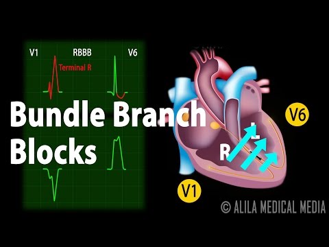
Bundle branch blocks happen when there is an obstruction in one of the bundle branches. The names "left bundle branch block" and "right bundle branch block" indicate the side that is affected. In a normal heart, the two ventricles are depolarized simultaneously by the two bundles and contract at the same time. However, in bundle branch blocks, the unaffected ventricle depolarizes first. The electrical impulses then move through the myocardium to the other side, resulting in a delayed and slow depolarization of the affected ventricle. This leads to a broader QRS complex, typically longer than 120 milliseconds, and a loss in ventricular synchrony. Left and right bundle branch blocks are diagnosed and differentiated by looking at ECG recordings obtained from the chest leads. The most useful leads for this purpose are V1 and V6, as they are best located to detect impulses moving between the left and right ventricles. In normal conduction, activation of the ventricle starts with the interventricular septum. Depolarization of the septum is initiated from the left bundle, going to the right toward V1 and away from V6. In left bundle branch block, there is a small positive deflection in V1 and a negative deflection in V6. The signals then move in both directions to the two ventricles, but as the left ventricle is usually much larger, the net movement is to the left, away from V1 toward V6. This corresponds to a negative wave in V1 and a positive wave in V6. In right bundle branch block, the initial septal activation is unchanged. The left ventricle depolarizes normally toward V6 and away from V1, producing a positive deflection in V6 and a negative deflection in V1. The impulses then reverse direction, spreading to the right ventricle. This results in a sub quint negative wave in V6 and a positive...
Award-winning PDF software


Video instructions and help with filling out and completing Who Form 8655 Bundles